
How Digistain Works?
Digistain® analyses the infrared spectroscopic signature of tumour tissue to provide recurrence-risk information
The Science Behind Digistain
Digistain utilises infrared spectrometry to analyse the composition of tumour biopsies, and estimate the risk of breast cancer recurrence. Infrared spectrometry is commonly used by chemists to deduce the chemical structure of large molecules. It is also used in various other fields of science and technology including chemical analysis, environmental monitoring, material characterisation, forensic analysis, and astronomical studies.
How Does Infrared Spectrometry Work?
Different chemical groups in a biomolecule absorb infrared light at very specific wavelengths. As the light passes through or reflects off the sample, certain wavelengths are absorbed, while others pass through. This absorption creates a unique pattern, known as an infrared spectrum, which is a graph showing the amount of light absorbed at each wavelength.
The peaks on the spectrum represent wavelengths where absorption occurs, corresponding to specific vibrations of chemical bonds within the molecule. By analysing these peaks, scientists can identify the functional groups present in a biomolecule. This allows them to determine its molecular structure and composition by acting as a molecular “fingerprint”, enabling precise identification of the biomolecule based on its unique vibrational characteristics.
By analysing wavelengths that are known to be absorbed by nucleic acids and cell proteins. This imaging approach enables Digistain to measure chemical concentrations associated with tumour malignancy, analyse tissue characteristics of cancer cells and quantify validated pathology markers correlated to tumour proliferation.
How Does the Digistain Test Work?
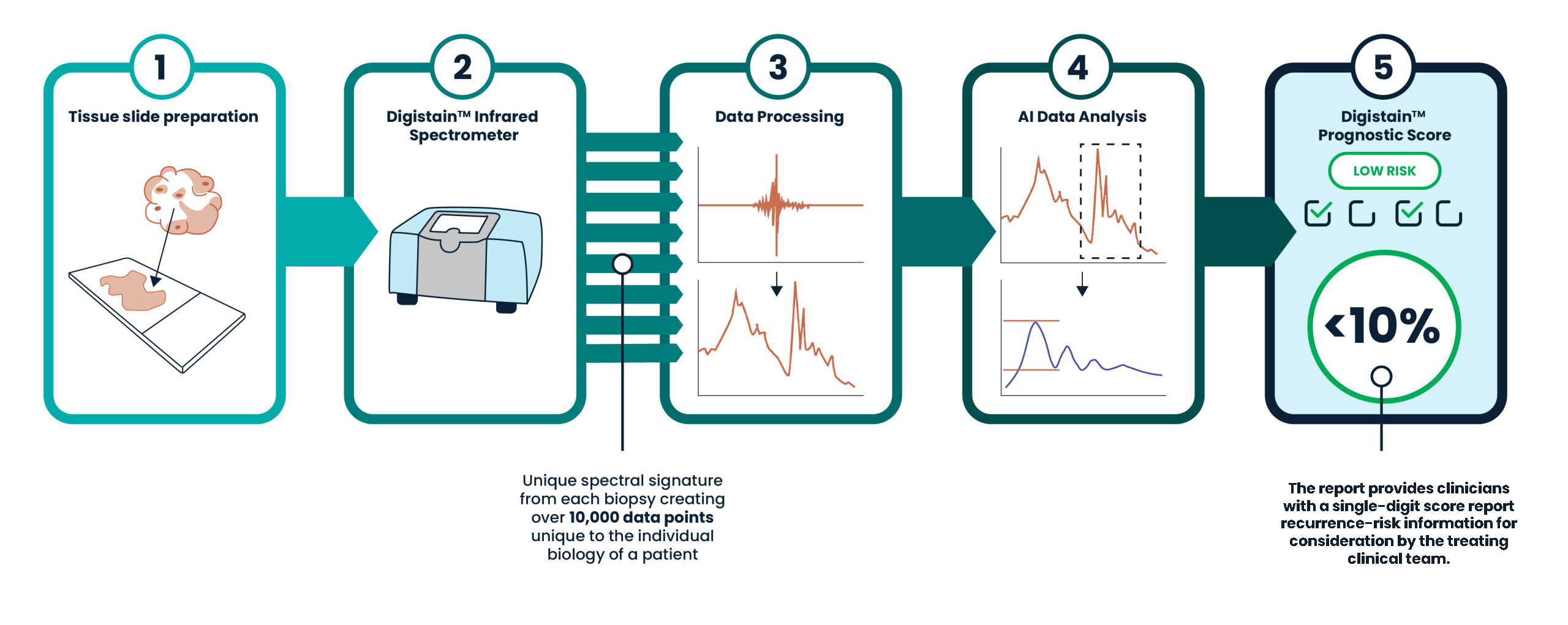
Using infrared spectrometry, Digistain captures a unique spectral signature from each biopsy and performs analysis on over 10,000 data points per biopsy to produce the Digistain Index.
Using this data, Digistain’s validated multivariate statistical model integrates that integrates key clinicopathological factors, such as age, tumour grade, tumour size, and lymph node status. It then identifies risk profiling patterns and computes a risk score – the Digistain Prognostic Score.
The report provides clinicians with a low/high result report recurrence-risk information for consideration by the treating clinical team.
H&E Stained


DIGISTAIN® Optical Scan
Image on the left shows traditional H&E staining approach. Image on the right shows Digistain approach with greater tumour contrast and detail enabling higher precision measurement
Subscribe to leading news and insights
To stay updated about Digistain’s developments, enter your details opposite. We promise only to send you important news.
Digistain Prognostic Score – Breast is an in vitro diagnostic test for professional use. The result is an estimate of the risk of recurrence and is probabilistic. It is used with, and does not replace, established clinico-pathological factors and clinical judgement. Treatment decisions remain with the treating clinician.